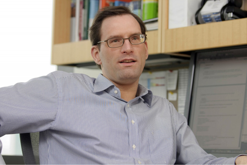
At Work: Radiologist and Nuclear Imaging Specialist Jan Grimm

Physician-scientist Jan Grimm works to develop innovative molecular imaging approaches for improved detection and monitoring of cancers. We spoke with him about his work in 2010.
Both my parents are scientists, so I was exposed to scientific minds from my early youth. By the time I reached high school in Hamburg, Germany, I chose biology and chemistry as the subjects of my advanced study. Even then, I was less interested in classical biology and more interested in cancer biology. I found it fascinating that there were these cells that were going haywire, and that the causes for these changes were still relatively unknown.
Around this time, my father, who is a zoologist, caught a tropical disease on a scientific expedition to Africa. He was initially misdiagnosed, which delayed treatment unnecessarily and caused additional complications. At some point, my mother brought his former, now retired doctor onto the case.
After examining a sample of my father’s blood under a microscope, he was able to correctly diagnose leishmania infection, a parasitic disease spread by the bite of sand flies. The experience made me realize how important the correct and factual approach is in medicine. It also inspired me to follow my interests and go to medical school.
The Benefits of Reading an X-Ray
In the final year of medical school at the University of Hamburg, every student had to do three clinical rotations: one surgical, one internal medicine, and one elective. I had always liked radiology, and I figured that knowing how to read an x-ray would always be useful later on in my career, so I chose it for my elective.
I ended up really liking radiology because I realized that it inserts you right in the middle of the entire patient-care process. For instance, working in the emergency room, you have the immediate treatment team made up of ER doctors, surgeons, and anesthesiologists.
As the radiologist looking at the scans, you’re always the first one to know what is wrong with a patient…and you’re having a positive impact on the patient’s recovery from the comfort of your chair. Also, I was fascinated by the technical aspect of the scan technology. These days, the scanners we use provide unsurpassed images of the inside of a living body, showing its function and physiology — now at the molecular level.
In Germany, once you’ve completed the coursework in medical school, you still have to write a thesis to get your MD degree. I ended up focusing on stem cell enrichment in bone marrow transplantation, using a specific method known as centrifugal elutriation. This thesis also led to my internship position.
First Taste of Molecular Imaging
Following an internship in bone marrow transplantation, I finally decided to pursue radiology as a career and applied for a radiology residency at the University of Schleswig-Holstein’s medical center in Kiel, Germany. From my bone marrow transplant work, I had an interest in imaging using antibodies coupled to iron oxide particles, which were used for cell depletion or enrichment. Without knowing anything about the emerging field of molecular imaging at that time, I suggested to the department chair during my interview that we could use these iron-oxide labeled antibodies as a specific and targeted contrast agent for MRI.
There was no radiology lab at Schleswig-Holstein when I started in 1997, so I had to find someone at the university to help me. Eventually, I located a professor of experimental oncology, Holger Kalthof, who had an antibody that had been used for tumor targeting but had not received any follow-up study. He liked the idea, and we started a collaboration — a collaboration that is still alive today.
It took us awhile to couple the iron oxide particles to the antibody, but we eventually were able to use it for imaging in mice. At the time, there were no animal-imaging magnetic resonance scanners available at the hospital, so we used the clinical MRI scanner at night, from to The resolution was not great, and we finally bought an MR coil specifically made for mice. This was the first molecular imaging I had ever done, and it was thrilling.
Going to the Source
At the same time that I was doing these early molecular imaging experiments, I began to do some interventional radiology work during my residency. While I found it very engaging, I knew that if I wanted to really learn more about the field of molecular imaging, I would need to go elsewhere.
And since I had been following the work of Ralph Weissleder, who is the founder of Massachusetts General Hospital’s Center for Molecular Imaging (and now also the director of the Center for Systems Biology), I knew that this was the place I had to go.
In 2002, I applied for and received a two-year research grant from the German Research Society to study with Ralph at Harvard. The work they were doing defined the field of molecular imaging. As a postdoctoral fellow, I worked on a number of interesting projects, including molecular imaging with MRI and iron oxide nanoparticles, activatable optical agents, and cell tracking with SPECT/CT.
In the fall of 2002, Ralph came to me and said that there was a faculty job available that he thought I should take. My original plan had been to complete the two-year research fellowship and then return to Germany for the typical university career. I discussed this offer with a lot of people — my mentor in Kiel, friends, colleagues, and my family — but I knew from the start that I really should take the job.
The Siren Call of the Clinic
As a result, after completing my postdoctoral fellowship, I accepted the faculty position as an instructor in the Department of Radiology at Harvard Medical School. I had a great time in Boston! In the first few years there, I was happy to be devoting all of my time to research. It was nice to have no clinical duties, no clinical meetings to attend. But then, as time went on, I started missing the clinical side of things. Even more surprising to me, I actually started dreaming about being back in the clinic.
After several years without clinical work — I was not yet licensed in the US — I realized that I either needed to take up at least a bit of clinical work again or risk losing my clinical skills completely. Therefore I decided that I needed to return, at least as part of my career, to the clinic, though I wanted it to be in a position where I could still pursue my molecular imaging work. To get an appointment as an attending at Harvard was difficult for various administrative reasons, so I started looking around at other academic research hospitals.
One of the people I contacted was Hedvig Hricak, the Chair of the Department of Radiology at Memorial Sloan Kettering. She emailed me back the following day and arranged to have me come down for an interview the following week. For me, Memorial Sloan Kettering was such an obvious choice, considering the amount and quality of translational research that is taking place here.
We agreed to an arrangement that would allow me initially to spend about 20 percent of my time in the lab and 80 percent in the clinic, so that I could get back up to speed clinically. This was a wise choice for me, since I’d never worked in a clinical setting in the United States (or in English!) before.
The Rewards of Working “Both Sides of the Street”
Working on the clinical side of things again was a real transition. There was a bit of a learning curve getting back up to speed in a field I had been out of for a number of years, but I started with a fellowship in body imaging and continued with a “fast-tracked” residency in nuclear medicine, which led to an appointment as assisting attending in the Department of Radiology.
In general, I enjoy working in both the clinic and the lab. It allows me to weigh what is relevant in the lab and what is possible in the clinic. It is important to see, firsthand, what clinical problems need to be addressed. Clinical work is also very helpful in resetting your mind and getting some distance from the research.
And the same is true in reverse. If you are stumped by a research problem, you can go to the clinic and use a completely different part of your mind. When you come back again to the lab, it helps you to refocus on the problem.
Bridging the Gap
My research involves using molecular imaging as an interdisciplinary approach that combines the knowledge of different fields to create innovative and unique new agents for various imaging applications. The ultimate aim of molecular imaging is to detect diseases earlier and with greater specificity. Most of today’s imaging agents — not including nuclear medicine agents — are nonspecific and are, at best, able to indicate physiological parameters such as perfusion and diffusion. There are largely no agents that specifically indicate the presence of, say, a specific antigen that is highly expressed on tumor cells or that is binding to a mutant kinase.
There is still a considerable disconnect between the advancements of modern medicine, which is moving into the molecular and designed world of biologicals (such as Gleevec), and the advancements in radiology, which are mainly driven by the equipment vendors and technical progress in scanner technology. An x-ray of the hand is basically done the same way as it was done 100 years ago.
Molecular imaging is aiming to bridge this gap by a) providing contrast agents that target specific molecular or cellular processes, and b) expanding the field of imaging modalities by, for example, introducing new imaging modalities like optical imaging into the clinic. This also aids in the development of drugs that are tailored to the specific needs of the patient. Identifying imaging agents capable of revealing the biological functions of their targets would significantly improve our ability to diagnose disease and also to monitor these novel therapies with the same agents.
My Current Research
My laboratory is interested in developing multifunctional targeted and activatable probes that are ideally not limited to one specific type of cancer. These agents are activated, or switched on, by their specific interaction with a dedicated target, which is usually an enzyme but can also, for example, be comprised of a certain DNA or RNA sequence.
Since these probes only produce a signal when their target is present and active, they have a very good signal-to-noise ratio. This means that they give very little or no signal in the absence of their target. Additionally, since one enzyme can activate many of these probes, there is also a considerable amplification of the signal. Many of these approaches are based on nanoparticles.
We are developing molecular imaging agents that seek to couple the unique magnetic, physical, and chemical properties emergent on the nanoscale with disease-relevant triggers. We are working with all the main imaging modalities — optical, MRI, PET, and CT, as well as with some ultrasound.
Just recently we were among the first few labs to explore a completely new imaging approach known as Cerenkov imaging of medical isotopes. This approach utilizes Cherenkov light induced by particle-emitting isotopes. (Particles traveling faster than the speed of light in water produce a shock wave equivalent to a sonic boom. This shock wave takes the form of blue light called Cherenkov light, named after the Russian physicist, Pavel Cherenkov, who first characterized it.)
Currently, we are also looking at prostate-specific membrane antigen (PSMA) as an imaging target. PSMA is highly expressed in prostate cancer tumors but also on the neovasculature of other tumors. Angiogenesis, defined as the formation of new blood vessels, is known to be critical for tumor progression and correlates with tumor aggressiveness. An imaging probe able to reflect the newly formed vasculature around tumors would therefore be a powerful tool to identify lesions, regardless of their type or origin.
I am striving to develop an interdisciplinary aspect to the research done in the lab. For the imaging agents we are working with, different expertise is needed in a variety of fields, such as chemistry, material sciences, molecular biology, radiology, and physics. Therefore it is a very diverse environment, one that creates ample opportunity to learn new things and to collaborate.


