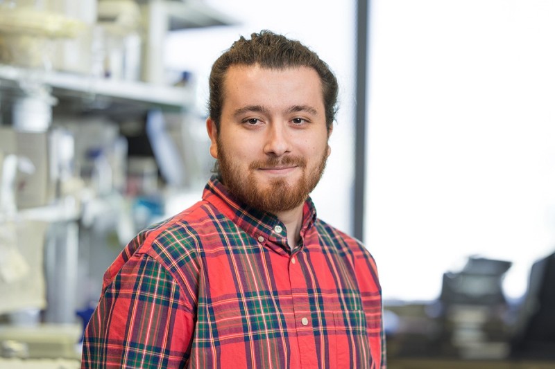
During my Ph.D. at Istanbul Medipol University (Turkey), my major focus was elucidating the molecular mechanisms of contraction after axotomy injury in dorsal root ganglion neurons using advanced microscopic and molecular techniques, and the establishment of the Advanced Imaging Laboratory and Cell Culture Laboratory (especially culture of primary neurons). I handled the microscopy systems, which included some of the most advanced setups from Zeiss such as a 2-photon microscope, LSM 780-800-880 (with fast airy scan) Confocal systems, a Spinning Disk Time Lapse system, a PALM microdissection and tweezer system, Axiozoom V16, GeminiSEM (SEM and TEM) and several stereo microscopes. In 2017, I finished my master’s degree in the field of Neuroscience, with a thesis titled “The effects of axotomy injury on membrane tension of dorsal root ganglion neurons”. I received a Ph.D. degree in the field of Biochemistry, and my thesis title was “The role of Hippo pathway in axolotl limb regeneration” in 2021. You can see my laboratory experiences below:
o Advanced Imaging Techniques
o Zeiss GeminiSEM (Both SEM and TEM)
o Zeiss LSM 780-800-880 Confocal Microscope
o Zeiss Micro Dissection and MicroTweezers Microscope
o Zeiss Time Lapse Confocal Microscope (Spinning Disk)
o Live Animal imaging and Manipulation (2-Photon Microscope)
o Live Cell imaging (including Ca++ and other ions imaging) and Manipulations
o Primary and Secondary Cell Culture Systems
o Dorsal Root Ganglion İsolation and Culturing from Mice and Axolotls
o in vitro Axotomy Model
o Measurement of Cellular Forces of Neurons
o Hippocampal and Cortical Neurons Culturing from Newborn Mice
o Mesenchymal Stem Cells İsolation from Human Bone Marrow
o Differentiation of Mesenchymal Stem Cells to Functional Neurons
o Sample Preparation for Scanning Electron Microscopy
o Axolotl Limb and Tail İnjury Models İmaging of Bones of Axolotls Using Micro-CT
o mRNA İnhibition of Axolotls Using Morpholinos Isolation of Certain Organs
o Several Cellular and Molecular Techniques
I am currently a Research Scholar at Dr. Gabriela Chiosis’ lab. My major focus is visualizing epichaperomes using several compounds. Our lab has discovered that context-dependent changes in protein networks within disease are executed by a restructuring of a fundamental functional protein group – the chaperome (a collection of chaperones, co-chaperones, and other factors) – into higher-order long-lived assemblies termed epichaperomes. Unlike chaperones, which fold and act through one-on-one dynamic complexes, epichaperomes mediate how thousands of proteins anomalously interact and organize inside cells, which aberrantly affects the function of protein networks, and in turn, cellular phenotypes. Epichaperomes have thus emerged as a conceptually innovative and translationally significant gateway to target, detect and control dysfunctional interactomes, now translated to clinics in cancer and Alzheimer’s disease, for both treatment and diagnosis. To advance our understanding of epichaperomes in cancer and learn important mechanistic insights, we develop chemical probes and methods for use in confocal (high resolution) and single molecule super-resolution imaging approaches.