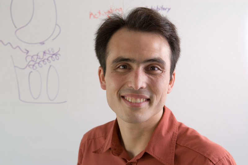
Immunologist Morgan Huse investigates intracellular signaling dynamics in lymphocytes. We spoke with Dr. Huse about his work in 2008, a year after he joined the Sloan Kettering Institute.
What first attracted me to science was that it allows you to know things with certainty, in a way that isn’t wishy-washy. My initial plan was to go to medical school. By sophomore year of college, friends of mine were already preparing for the MCAT, and I thought to myself, “What a drag!” It struck me that if I was turned off by a standardized test, then medical school was probably not for me.
I did, however, enjoy my collegiate biology research experience. As an undergraduate, I worked as a research assistant in Stephen Harrison’s lab, which focused on the three-dimensional structures of viruses and signaling molecules. I enjoyed this work considerably, in part because as a structural biologist, you can ask very specific questions and get concrete answers.
In addition, the idea that we are all large assemblies of tiny molecular machines was fascinating to me. In some ways, I’ve never gotten beyond this fascination with basic molecular biology and biochemistry.
A New York Welcome
In considering different graduate schools, I decided on the Rockefeller University for a combination of reasons. It was a very dynamic place to be doing research in structural biology, with scientists like John Kuriyan, Stephen Burley, Rod MacKinnon, and Seth Darst all working in close proximity. It also seemed like the sort of place where you could write your own ticket as a graduate student. College was very enjoyable for me, but I was tired of being assessed every three to four weeks.
Plus, I was excited to move to New York. I was actually born in Manhattan, but my family moved overseas when I was just three months old. I spent a good deal of my childhood in Asia: Bangkok, Seoul, and Tokyo, to be specific, which are very interesting cities in their own right. However, I always felt an affinity for New York, perhaps because it was my birth city, perhaps because when you mention New York outside of the USA, people know what you’re talking about.
I find the diversity, density, and frenetic pace of life in New York to be liberating. Just to give an example, some of the best biomedical research in the country is performed at Sloan Kettering Institute, Rockefeller, and Weill Cornell Medical School, and yet if you walk three blocks west of here, most people couldn’t care less. I love that about New York. It helps keep things in perspective.
Research Focus at Rockefeller
At Rockefeller, I became interested in the molecular mechanisms of signal transduction, specifically how phosphorylation can switch a signaling protein from an “off” state to an “on” state. The way this often works is by allostery, where a portion of the protein is autoinhibitory in the dephosphorylated state, but becomes stimulatory upon phosphorylation.
I did my thesis work in John Kuriyan’s lab, where I studied the regulation of the type I TGF-β receptor (TβR-I), a serine/threonine kinase that is important for development and homeostasis. In doing so, I collaborated extensively with Joan Massagué, [then] Chair of the Cancer Biology and Genetics Program here at Sloan Kettering, and a leader in the study TGF-β signaling.
TβR-I is regulated by phosphorylation of the GS region, a little stretch of amino acids that’s just N-terminal in the kinase domain. It had been shown in Joan’s lab that you need four to five phosphate groups in the GS region in order to fully activate the kinase. I became very interested in how this process actually works. We solved the structure of unphosphorylated TβR-I, which included the GS region and the kinase domain, in complex with another protein called FKBP12, which binds to TβR-I and stabilizes its inhibitory conformation.
We found that in the dephosphorylated state the GS region maintains the kinase in an inhibited conformation that is inconsistent with catalytic activity. That inhibited conformation is capped and stabilized by FKBP12. The structure showed us how the protein was turned off in the dephosphorylated state, but unfortunately it gave us little information about how multiple phosphorylation of the GS region activated the enzyme.
In order to study that transformation as biochemists and structural biologists we needed to make multiply phosphorylated TβR-I. This protein proved exceedingly difficult to produce by standard biochemical methods, so we developed an approach that involved synthesizing the phosphorylated GS region separately and then attaching it to the rest of the protein using the native chemical ligation reaction, a process that allows you to stitch together large proteins from smaller peptide segments.
The production of this semisynthetic protein was done in close collaboration with Tom Muir, a synthetic protein chemist at Rockefeller who participated in the initial development of the native chemical ligation reaction.
The ability to produce multiply phosphorylated TβR-I allowed us to perform biochemical experiments to directly investigate the activation process. We were able to show that phosphorylation of the GS region transforms it from a binding site for the inhibitor FKBP12 into a binding site for transcription factor Smad2, the physiological substrate of TβR-I. These results explained the central role of phosphorylation in TβR-I signal transduction.
Approaching Postdoctoral Research
After completing my PhD, I was urged by a number of people to round out my education and really test myself by moving into a different field. I took them seriously and ended up moving to Stanford University, where, after a bit of wandering, I ended up in Mark Davis’ lab. The Davis lab studies T cell activation and the T cell receptor (TCR) signaling network using a combination of biochemical and biophysical approaches that incorporate high-resolution imaging technology.
I found both the approach and the subject matter very compelling, in part because of my long-standing interests in cell signaling and molecular assemblies and, in part, because as a structural biologist I am very image oriented.
I worked on a couple of different things in the Davis lab. My first project focused on how T cells communicate with other components of the immune system. T cells recognize little pieces of foreign pathogen (called antigens) presented on the surface of other cells. This recognition event activates the T cells, which respond by forming a tight contact, called an immunological synapse, with the antigen-presenting cell (APC). Then, the T cell secretes a number of different messenger molecules, called cytokines, which trigger responses in the surrounding cells.
We discovered that there are two directionally distinct cytokine secretion pathways in activated T cells. The first pathway targets one group of molecules to the immunological synapse, while the second releases a different set of factors in a multidirectional manner. Thus, the T cell appears to have two modes for intercellular communication: a “private” system for the delivery of specific signals to the APC and a “public” pathway used to trigger inflammation and to recruit other cells.
For my second project, I used my background in peptide chemistry to develop a new way to study T cell receptor (TCR) signaling dynamics. First, we used a little bit of synthesis to prepare a “photocaged” ligand for the TCR that is nonstimulatory until you irradiate it with UV light. Then, we used this reagent to trigger T cell signaling responses in the context of an imaging experiment.
This involved coating a glass coverslip with the photocaged ligand, allowing T cells to land on top of it, and then using a UV laser to create small regions of stimulatory ligand beneath individual T cells. This methodology allowed us to monitor early T cell signaling events with unprecedented spatial and temporal resolution.
For example, we were able to show that tyrosine phosphorylation of an early signaling component, the LAT adaptor protein, takes place in four seconds. Precise kinetic measurements like this will allow us to address some of the outstanding questions in T cell signaling, such as how T cells can be both sensitive and exquisitely selective for their cognate ligands.
Our studies also revealed that certain downstream signaling events, such as the influx of calcium into the T cell, desensitized within seconds of TCR activation, while others, such as LAT phosphorylation, remained sensitive to repeated stimulation. Based on these results, we concluded that different signaling events play distinct signal processing roles within the TCR signaling network.
This second project was particularly satisfying to me for two reasons. First, I was able to merge my interests in protein chemistry and intracellular signaling. Second, this methodology gives us a way to ask very specific biochemical questions in the context of a complex cellular response, essentially to do quantitative biochemistry in single cells. What’s more, there’s no reason that the approach couldn’t be adapted to study any number of interesting signaling systems.
Why SKI?
I stayed at Stanford until 2007, when I decided it was time to start my own lab. I was looking all over the country at a wide range of places — universities with undergraduates, hospitals, institutes, etc. The idea was that the search itself might help me determine what my interests were. Looking for a job is a very educational process. You end up doing a lot of thinking about what’s really important to you.
I chose Sloan Kettering Institute for a number of reasons. Working across the street at Rockefeller, I had always been impressed by the quality of the people and the work at SKI. In addition, there’s a lot of energy in the Immunology Program right now and we’re recruiting a number of young faculty, which gives the program this feeling of momentum.
I suppose what sold me was the idea that I could be part of something new and dynamic. Finally, the support for research at SKI is just incredible, especially in this day and age, with the NIH situation being the way it is. In coming here I knew that the institution would be behind me. I couldn’t be sure of that in a lot of other places.
Research Goals and Laying Claim to a Little Piece of the World
What we are trying to do in the lab is to combine synthetic chemistry and protein engineering with imaging in order to answer questions about lymphocyte signaling. Lymphocytes such as T cells, B cells, and NK cells play crucial roles in the immune response, and the signaling pathways that govern their behavior have been the subject of intense research for a number of years.
Most of what we have done, though, is genetic or biochemical in nature. These approaches are good at identifying important signaling molecules and establishing pathway connectivity. However, they aren’t as good at revealing other aspects of the signaling network — the kinetics, the localization, the dynamics, and the way the responsiveness of the network changes over time.
We want to get at this information by using controlled amounts of recombinantly expressed, purified ligands to trigger lymphocyte signaling responses in the context of an imaging experiment. We will use chemistry and protein design in order to give ourselves an added element of control in these studies.
The application of photocaging will give us the ability to control where and when we stimulate a specific receptor. In addition, the design of receptor ligands bearing specific fluorescent groups will allow us to correlate the amount of stimulation with a specific intracellular response, and in this way probe sensitivity.
I am very excited to be here at SKI. Many scientists, myself included, get into research because they want to explore their own little piece of the world. Now I have the opportunity to do that, in the way that I see fit. Not a lot of people can say that. I don’t know how else to say it, but I am really glad to be here. I hope I don’t blow the opportunity.

