Sexual reproduction is like shuffling the genetic cards. Not only are you a blend of the genetic attributes of your parents, but the sperm and egg that came together to make you were themselves the product of a genetic shuffling called meiosis.
Each time sperm and egg form from precursor cells, the chromosomes inside them are broken at hundreds of spots along their length. Maternal and paternal chromosomes swap segments before these breaks are resealed, and the rearranged chromosomes are then divvied up into sperm or eggs. This shuffling of genes between chromosomes is one of the main reasons why you look similar, but not identical, to your siblings.
The key molecular scissors that makes these breaks is called Spo11, a protein that Sloan Kettering Institute molecular biologist Scott Keeney discovered in 1997 when he was a postdoc at Harvard. For more than two decades, Dr. Keeney has been focused on understanding how Spo11 accomplishes this feat. (His enthusiasm for the endeavor is suggested by his Twitter handle: Spo11Rulz.)
A long-standing mystery about Spo11 is how its cutting activity is regulated so that it only makes cuts when and where it’s supposed to.
“Spo11 is dangerous,” Dr. Keeney explains. “It makes a very dangerous type of DNA damage called a double-strand break, and it wouldn’t really make sense for a cell to just turn loose this enzyme to cut anywhere in the DNA.” A double-strand break is when both sides of the DNA helix are broken.
For many years, unraveling this conundrum was hampered by scientists’ inability to isolate Spo11 from the welter of other molecules in the cell. “We tried really hard in the early days and completely failed,” Dr. Keeney admits. Without a pool of pure protein to work with, they had to rely on other measures of Spo11’s activity.
It wasn’t until a postdoc in Dr. Keeney’s lab, Corentin Claeys Bouuaert, came along that the lab was finally able to isolate Spo11 and the proteins with which it interacts. Dr. Keeney credits Dr. Claeys Bouuaert’s extraordinary skills as a biochemist for cracking the nut. The team published those results in Nature Structural and Molecular Biology in January 2021.
Dr. Claeys Bouuaert now has his own lab at the Louvain Institute of Biomolecular Science and Technology in Belgium, but he and Dr. Keeney continue to collaborate, most recently on a project whose results were published March 17, 2021, in Nature. The findings build on the previous paper and cap a more than 20-year intellectual journey.
From Complexes to Condensates
With Spo11 and its partners finally purified, the scientists could ask how all these proteins interact with one another by studying them in the controlled environment of a test tube. The results were surprising.
“We knew there were 10 proteins total in the cutting process — nine other proteins in addition to Spo11,” Dr. Keeney says. “Based on how other cellular enzyme complexes are thought to work, we assumed that these 10 proteins functioned as a discrete, functional unit. But what we show in this paper is that it’s not that way at all.”
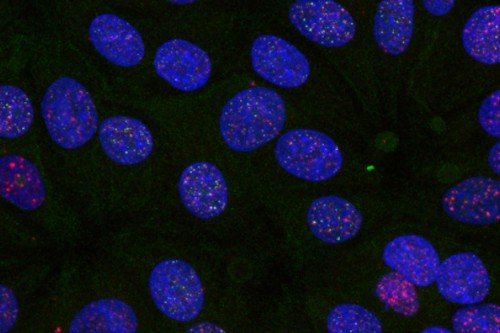
Instead, the proteins form much larger structures: massive assemblies of proteins piled along the length of a chromosome. And it’s at these large hubs — what are termed “biomolecular condensates” — where the double-strand break activity is concentrated.
If you imagine DNA as a 500-foot long piece of string, then the Spo11 condensates would be like huge buoys bobbing along it every few feet or so.
The discovery helps to explain another perplexing aspect of Spo11’s activity. “For all its importance, Spo11 by itself is actually a pretty crappy enzyme,” Dr. Keeney says. “It just doesn’t work very well.”
The massive Spo11 condensates that the team discovered explain why this isn’t a problem — and in fact is an advantage. Even though each Spo11 enzyme is only weakly effective, together they assemble into a very good cutting machine. And because it’s all concentrated in one place as well, the cell can control the placement of cuts so things don’t get out of hand.
A Shift in Perspective
For Dr. Keeney, the discovery was pleasantly disorienting. “It’s like looking at one of those optical illusions where at first it just seems like noise, but if you stare at it long enough, all of a sudden something emerges from the noise.”
He says the new perspective gives the team new questions to ask, new roads to follow.
Besides providing insights into the process of meiosis, there are also connections to cancer. Spo11 belongs to a class of enzymes called topoisomerases, whose main job is to cut DNA in order to relax its supercoiled structure. Think of a full-length DNA molecule as being like an old-fashioned telephone cord that coils up on itself into a knot. By briefly cutting and resealing the DNA strands, topoisomerases relax the coil and allow important DNA business to take place along its length. Certain chemotherapy drugs specifically inhibit the resealing function of topoisomerases, which causes DNA breaks to go unrepaired so that the cells die.
The new discoveries wouldn’t have been possible without the help of Memorial Sloan Kettering’s core facilities, which afford MSK researchers unbridled technological support for conducting novel experiments. In particular, the Microchemistry and Proteomics core, the Molecular Cytology core, and the Integrated Genome Operation were essential, Dr. Kenney says.
The project was a collaboration with SKI structural biologist Dinshaw Patel and funded in part by an MSK Basic Research Innovation Award. Dr. Keeney is a Howard Hughes Medical Institute Investigator and received support from that organization as well.


