How Computational Biology and Artificial Intelligence Are Changing Cancer Care
The microscope. The petri dish. The X-ray. A handful of tools have radically changed the practice of medicine and biomedical research.
Although easy to overlook, the computer microchip, with its layers of silicon and engineering wizardry, is without a doubt one of the most important.
Today, as the instruments used in the lab and the clinic become more sophisticated, computation plays an increasingly essential role in scientific discovery and in improving patient care.
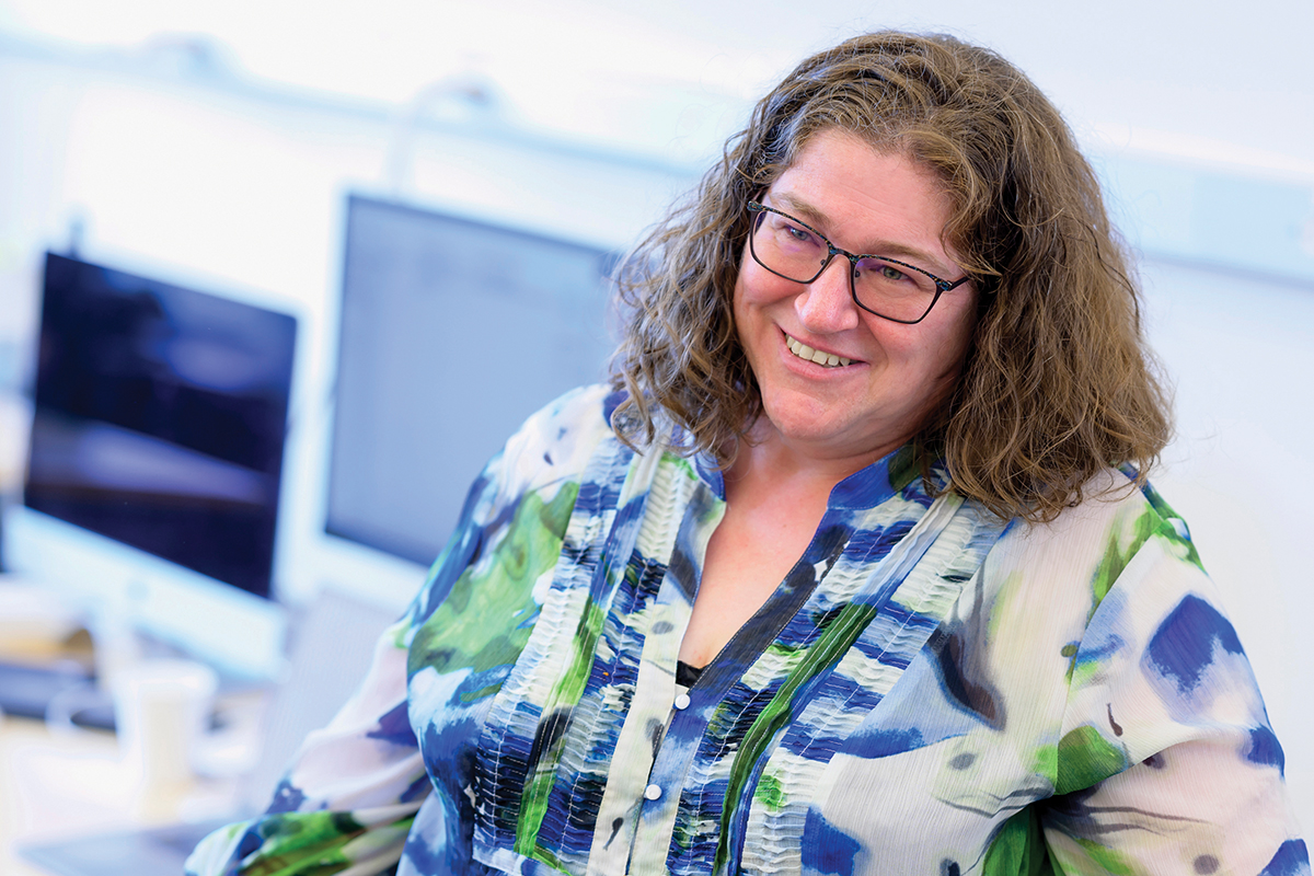
“Biology is really becoming an information science,” says Dana Pe’er, PhD, an investigator at Memorial Sloan Kettering Cancer Center (MSK) and Chair of the Computational and Systems Biology Program at MSK’s Sloan Kettering Institute.
And what sets MSK apart is the ability for computational and cancer experts to work together as partners to illuminate human biology and improve treatment outcomes.
Dr. Pe’er’s research group, for example, recently developed a computational tool — dubbed Spectra — to help analyze research done with a technique called single-cell sequencing. The technology is akin to taking an orchestra and isolating individual musicians from it, or groups like the woodwinds.
By guiding data analysis in a unique way, the Spectra algorithm will provide new insights into the complex interplay among thousands of cells, including those that are critical to helping today’s groundbreaking immunotherapy treatments work for more people.
“Interpreting this type of data is incredibly complex and rife with many statistical pitfalls,” Dr. Pe’er says. “Spectra both finds patterns that are too complex for a human to possibly identify and also easily finds patterns that would take researchers months using other methods.”
AI and Cancer Care
The importance of computation isn’t limited to the laboratory. MSK is a world leader in the clinical use of home-grown artificial intelligence (AI) models, notes Joseph Deasy, PhD, Chair of MSK’s Department of Medical Physics. For example, AI is being used routinely to improve the efficiency and quality of radiation therapy treatment planning.
MSK researchers, led by computer scientist Harini Veeraraghavan, PhD, have developed AI methods — trained on MSK imaging data — that can accurately identify healthy tissues that can be spared while zeroing in on tumor tissues. Thanks to this research, the approach now covers more than 40 tissue types and has been used for over 6,000 cancer treatments, making it a great example of the positive impact AI is already starting to have in medicine.
What’s more, because AI programs can be trained to adapt and improve over time, they perform in ways far superior to traditional computing approaches.
How Data Is Improving Patient Care
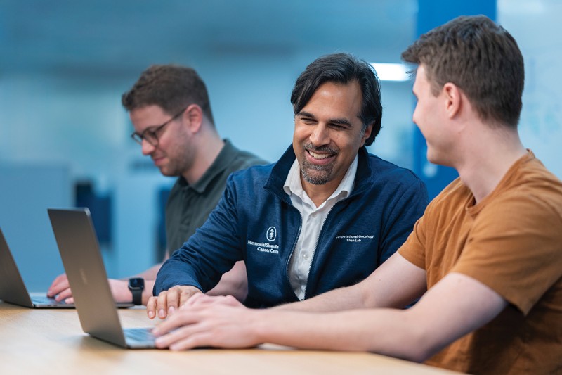
And it’s not just AI that is making a difference. Harnessing the power of large data sets is central to improving cancer diagnosis and continuously advancing patient care, says Sohrab Shah, PhD, who heads MSK’s Computational Oncology Program. Moreover, MSK is poised to extend its leadership in the field, thanks to the Halvorsen Family Foundation’s transformational $25 million gift establishing The Halvorsen Center for Computational Oncology. Among the top priorities for the center are investigating tumor evolution and drug resistance, immuno-oncology, and AI-powered next-generation personalized medicine.
This includes the river of data created during the process of providing care to patients with cancer — an electronic record that includes everything from their age, ethnicity, and gender to the genetic mutations driving their cancer and measurements of how much a particular medicine shrank their tumors. Every week, some 10,000 imaging scans are done at MSK — each rich with electronic data.
“The primary purpose of gathering all this information is to treat each patient properly and effectively,” Dr. Shah says. “But all this information has enduring value by creating a large data set from which patterns can be analyzed for the benefit of future patients.”

Dr. Shah helped lead research, for example, that performed a variety of advanced analyses on samples from patients with high-grade serous ovarian cancer — one of the most challenging cancers to treat.
The team uncovered several mechanisms that help explain why ovarian cancers have been resistant to immunotherapy, finding the disease is even more complex than previously thought. There are profound differences in the ability of different types of ovarian cancer to evade the patient’s immune system. Their findings were published in Nature, one of the top scientific journals in the world.
“Uncovering these mechanisms that are driving resistance to therapy creates an opportunity to find ways to improve treatments,” Dr. Shah says. “It also will lead to better methods for both detection and prevention.”
Harnessing the power of computation is helping in other ways, too. AI technology has already sped data collection and cut scan times in half in recent years, says Lawrence Schwartz, MD, Chair of MSK’s Department of Radiology.
“In many settings, a healthy woman’s mammogram is read for a second time with an AI technology to make sure that nothing was missed,” he says. “But we’re certainly just at the beginning of the journey to understand what AI can do.”
Dr. Deasy holds the Enid A. Haupt Chair in Medical Physics.
Dr. Norton holds the Norna S. Sarofim Chair in Clinical Oncology.
Dr. Pe’er holds the Alan and Sandra Gerry Endowed Chair and is a Howard Hughes Medical Institute Investigator.
Dr. Shah holds the Nicholls-Biondi Chair.