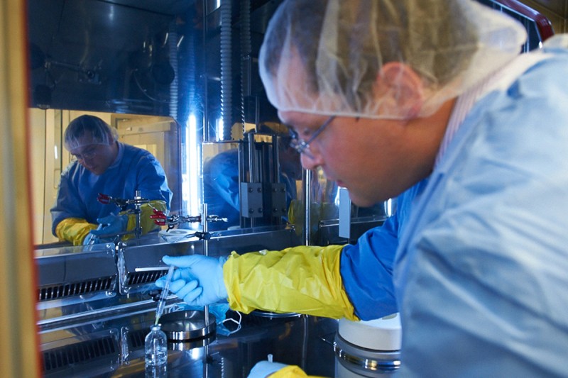
Radiopharmacist Serge Lyashchenko preparing a reagent in the cyclotron facility.
Memorial Sloan Kettering has taken a leap into the future with the launch of a new cyclotron, a type of particle accelerator that will be used to produce radioactive molecules for PET imaging of cancers. The 44,000-pound instrument and the production facility built around it are expected to change the way our patients are diagnosed and treated by allowing doctors to examine and target tumors with increased precision.
“This is truly a cutting-edge facility and one of the largest of its kind in the world,” says radiochemist Jason S. Lewis, who runs the cyclotron facility and also directs the recently established Center for Molecular Imaging and Nanotechnology.
Affectionately nicknamed Dorothy, the machine was inaugurated during a festive ceremony on May 22. Speaking at the event, President and CEO Craig B. Thompson marveled at the tour de force of engineering and logistics required “to actually drop a 20-ton [cyclotron] through the middle of the hospital one weekend” in November and successfully install it in the basement while keeping the hospital fully operational.
At the same ceremony, Physician-in-Chief and Chief Medical Officer José Baselga noted that the cyclotron is part of a wide-ranging effort “to build the best cancer center in the world in precision-based medicine.”
“We have tremendous insights in biology. We have the capacity to produce the best therapies,” Dr. Baselga said. “And now, with this new facility, we will have the capacity to interrogate tumors and really know what’s going on in individual people’s cancers.”
Hot Tools for Medicine and Research
PET imaging makes it possible to watch biological processes play out in tumors or normal tissues using radioactively labeled imaging agents. “The PET scanner is our molecular microscope,” explains physician-scientist Wolfgang Weber, who heads the Molecular Imaging and Therapy Service. “It allows us to localize molecules in the body of a patient and measure their concentrations with amazing sensitivity. The PET scanner is literally counting individual atoms.”
The method can be used to assess whether individual patients will benefit from a drug, monitor the drug’s effectiveness, or study its mechanism of action in a clinical trial. Other types of PET scans are used to diagnose and stage cancers by measuring tumors’ glucose uptake. In addition, PET is a vital tool for lab studies into the basic biology of cancers.
Radioactive isotopes — the components of PET agents that make molecules visible in a patient — are produced in a cyclotron by hitting a nonradioactive molecule with a charged particle flying at extremely high speed, accelerated in a powerful magnetic field.
Hospital cyclotrons produce almost no radioactive waste, and patients who undergo PET imaging are exposed to very low doses of radiation.
A Bit of Cyclotron History
In 1967, Memorial Sloan Kettering was the first hospital in the country to install a cyclotron for nuclear medicine. “It was called Betsy and served us faithfully for almost 40 years,” recalls Hedvig Hricak, who chairs the Department of Radiology. “When old Betsy was finally decommissioned, she was partially held together with duct tape and Krazy glue!”
Betsy’s successor, a facility still in use, was set up on Manhattan’s East 72 Street and is shared jointly by Memorial Sloan Kettering and Weill Cornell Medical College. “But with the rapid development of cancer treatments and diagnostic procedures that involve sophisticated forms of image guidance, our nuclear medicine program soon outgrew the capacity of the 72th Street facility,” Dr. Hricak says. “With the new cyclotron facility we will be able to fully capitalize on our strength in radiochemistry, nuclear medicine, and molecular imaging and monitor and study cancers in unparalleled detail.”
Instant Drug Manufacturing
There is another reason a new cyclotron was needed within Memorial Hospital: Our researchers are creating new medical technologies that require radioactive drugs to be produced and delivered to patients very quickly.
“For example,” Dr. Lewis says, “the new facility will enable us to make carbon-11. This isotope can be used to produce hundreds of highly selective PET agents, including some of the best markers available for monitoring prostate cancer and some types of glioma brain tumors.”
But using the isotope in the clinic is challenging due to its short half-life — about 20 minutes — which is the time it takes for a freshly made batch of the isotope to decay until half of it is gone.
“While the patient waits in the scanner, we make the isotope, take it off the cyclotron, couple it with a tracer or drug molecule, sterilize the product, and perform nine different tests to ensure optimal quality and safety,” Dr. Lewis says. The compound is then propelled to the imaging suite via one of seven pneumatic tubes that connect the cyclotron facility with clinical areas of the hospital. For carbon-11 and a number of other isotopes, this whole process must be done within minutes.
“At Memorial Sloan Kettering we have developed more than 30 promising new PET agents,” Dr. Lewis notes. “And some are being used worldwide to translate new drugs into the clinic.”


