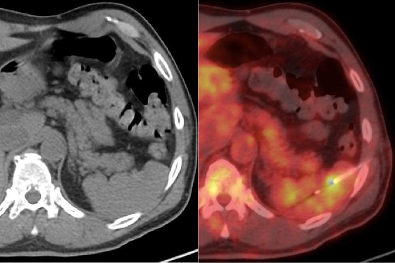
These images show a needle biopsy of a spleen in a patient with lymphoma. In the regular CT image (left), the tumor and needle are difficult to see. In the PET/CT image, both are visible, demonstrating the value of this technology.
Imaging technologies enable physicians and researchers to look deep within the human body. These tools — which include older techniques like x-rays and ultrasound as well as advanced methods like PET-CT and specialized forms of MRI — are advancing the understanding and treatment of cancer.
Interventional radiology is the name for the medical specialty that uses these technologies to perform procedures in patients. The operations can be done using nothing more than very small incisions in the body. These techniques are known as minimally invasive procedures. In the setting of cancer, these methods are commonly used to perform biopsies and treat tumors.
Until recently, patients under the care of Memorial Sloan Kettering interventional radiologists needed to travel to Manhattan for treatment. With the opening of two state-of-the-art outpatient centers — MSK Westchester in 2014 and MSK Monmouth in 2016 — and the expansion of MSK Commack, patients are now able to have many of these visits and procedures closer to home.
“Extending interventional radiology services to the MSK regional sites provides a convenient way for patients to benefit from our expertise in the communities where they live,” says Robert Siegelbaum, Director of Interventional Radiology at MSK Commack.
Image-Guided Biopsies
Biopsy is one of the most common procedures performed by MSK’s interventional radiologists. The technique utilizes a special needle that is guided to the site of the tumor using imaging such as ultrasound, MRI, or CT. It’s less invasive than open surgical biopsy and also allows samples to be obtained from areas that otherwise would not be easy to reach, such as the liver, pancreas, and bone.
Needle biopsies are typically done with local anesthesia applied to the skin and intravenous sedation. Most biopsy procedures can be performed at MSK’s outpatient sites, and are typically completed in less than an hour.
At Memorial Hospital, a specialist called a cytologist reviews biopsy samples while the patient is still on the procedure table. This enables a very high rate of sampling accuracy and decreases the need for repeat biopsies. Thanks to a specialized robotic microscope that can send images to doctors in Manhattan, patients who undergo needle biopsies at MSK’s outpatient sites are able to have their biopsies reviewed in the same way.
“Using this state-of-the-art equipment, a cytologist at Memorial Hospital is able to look at the sample in real time,” Dr. Siegelbaum says. “They can tell us right away if the tissue we obtained will likely provide a diagnosis for our patients.”
Placement of Ports for Chemotherapy or Blood Draws
Another procedure available to many patients at MSK’s outpatient sites is the placement of implantable ports or external catheters for administering chemotherapy and drawing blood.
Placing the catheters with ultrasound and fluoroscopy (real-time x-rays) imaging allows the interventional radiologist placing the device to ensure that it is positioned optimally.
“Imaging guidance is integral to success,” says Lynn Brody, an interventional radiologist who practices at MSK in Manhattan and Monmouth. “Optimal placement helps maintain good function and makes things easiest for the patients, as well as for the nurses and oncologists providing their therapy.”
Minimally Invasive Treatments
Interventional radiologists also provide a number of minimally invasive treatments to destroy tumors or stop their growth. These procedures are performed at Memorial Hospital in Manhattan.
With ablation, interventional radiologists use imaging technology to guide a special needlelike applicator to the site of the tumor. The needle produces a zone of extreme heat or cold that destroys the tumor.
With embolization, interventional radiologists use a tiny catheter inserted through the artery in a patient’s groin to deliver small beads that block the blood vessel supplying nutrients to the tumor. Embolization is most commonly performed to treat certain types of liver tumors.
Embolization and ablation are alternatives to surgery in some patients. “These procedures may be repeated several times over many years and are sometimes used in combination,” Dr. Brody explains. “We hope to control the tumors we treat well enough to significantly extend survival, even in cases in which it is not possible to cure the patients of their disease.”
Although these more complex procedures still need to be done in Manhattan (because an overnight hospital stay is typically required) patients are able to have their clinic consultation, lab work, and follow-up care at the outpatient sites in Westchester, Long Island, and New Jersey.
“Everything can be planned and set up from the regional sites,” Dr. Brody says. “Patients generally don’t have to come to the hospital until it’s time for the actual procedure.”


