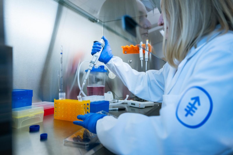
New research from Memorial Sloan Kettering Cancer Center (MSK) develops a new tool for studying mitochondrial DNA deletions; uncovers mechanisms of histiocytosis-associated neurodegeneration; and investigates how mutant stem cells co-opt regeneration to make tumors grow.
A new tool for studying mitochondrial DNA deletions
The laboratory of molecular biologist Agnel Sfeir, PhD, at MSK’s Sloan Kettering Institute, studies how the body repairs broken DNA and how mismanagement of this process can lead to diseases. This includes mitochondrial DNA (mtDNA), genetic material located in the mitochondria, the energy-producing organelles dispersed throughout the cell’s cytoplasm.
When mtDNA undergoes large-scale deletions, it can result in severe disorders that involve muscle weakness, neurologic degeneration, and heart defects. Now Dr. Sfeir’s lab has developed a novel method for engineering specific mtDNA deletions in human cells. They collaborated with the Single Cell Analytics Innovation Lab (SAIL) at MSK, as well as with the labs of computational biologist Dana Pe’er, PhD, and cell biologist and former MSK President and CEO Craig B. Thompson, MD, to develop the technique, which provides a powerful tool to model disease-associated mtDNA deletions in different cell types.
Deleting genes in mtDNA to understand their function is much more difficult than doing so in nuclear DNA. The main reason is that the mitochondria have no repair mechanism to join broken DNA strands back together — the broken DNA is degraded. To overcome this hurdle, the research team used a “cross-kingdom” approach. (Cross-kingdom means organisms from different biological kingdoms.)
From bacteria, they borrowed a DNA joining mechanism — called end joining (EJ) machinery. They combined this with human enzymes that can be used to precisely modify DNA sequences. To demonstrate proof of concept for the research tool, they generated a panel of cell lines with more than 3,500 base pairs of mtDNA deleted.
The cells had varying proportions of normal (wild type) and mutated (or deleted) mitochondrial DNA within each cell. (Each cell contains multiple mitochondria, and each mitochondrion has multiple copies of mtDNA.) They found that mitochondria had a critical threshold for mtDNA deletions. The organelles could lose up to 75% of the genetic information before serious, disease-causing problems arose.
“This is a valuable resource for understanding the impact of mtDNA deletions on diseases,” Dr. Sfeir says. “The cross-kingdom approach worked beautifully, already providing important insights into how cells manage mtDNA deletions. It will help with therapeutic strategies — for example, is there a way for us to help cells keep mtDNA deletions below the critical threshold? That could help with fighting these diseases, for which there are currently no cures.” Read more in Cell.
MSK researchers uncover mechanisms of histiocytosis-associated neurodegeneration
Langerhans cell histiocytosis (LCH) and Erdheim-Chester disease (ECD) are rare disorders characterized by clonal myeloid cell abnormalities — referring to an overgrowth of abnormal myeloid blood cells originating from a single mutated cell. These diseases are not only associated with neurodegeneration but also a higher incidence of blood cancers and solid tumors.
A new study led by an international team of researchers investigated the underlying mechanisms of neurodegeneration in LCH and ECD. The team — led by MSK research fellow Rocio Vicario, PhD (now at Calico Life Sciences), and overseen by senior author Frederic Gesissmann, MD, PhD, an immunologist at MSK’s Sloan Kettering Institute — discovered that cells carrying a mutation in the BRAF gene accumulate in specific regions of the brain and drive the neurodegenerative process. These mutated cells, known as inflammatory microglia, were found in the brainstem, cerebellum, and hippocampus.
Importantly, the study revealed that early signs of the diseases can be detected years before clinical symptoms appear, indicating the existence of an early stage of degeneration that could potentially be targeted for therapeutic interventions. In mouse models, depleting the inflammatory microglia during this early stage showed promising results — reducing inflammation, protecting neurons, and improving overall survival. Read more in Neuron.
How mutant stem cells co-opt regeneration to make tumors grow
Tumors have long been thought to behave much like wounds that do not heal. New research from the lab of stem cell biologist Joo-Hyeon Lee, PhD, and colleagues at other institutions has revealed new insights into the mechanisms that underpin the striking parallels between normal regeneration and the development of tumors, including how lung stem cells with oncogenic mutations can hijack regeneration for tumor growth.
The researchers examined the trajectories of alveolar stem cell fates carrying the KRAS-G12D mutation, which is common in lung adenocarcinoma, a leading cause of cancer-related mortality worldwide.
The researchers discovered that a specific subgroup of these mutated stem cells has an enhanced capacity to generate tumors. The KRAS-mutant stem cells initially adopt a regeneration program during tumor initiation. However, dysregulated feedback mechanisms lead to sustained mutant NF-kB activation. This enables frequent reversible cell state transitions that drive tumor expansion and prevent normal cell differentiation.
“Our findings shed light on lung adenocarcinoma evolution and could help pave the way for new therapeutic strategies,” Dr. Lee says. Read more in Cell Stem Cell.