The composition of malignant tumors is incredibly complex. They contain not only cancer cells but also dozens of other cell types, such as supportive tissues, fat, and many kinds of immune cells. These other cells interact with the cancer cells and one another to influence how tumors behave.
To get to the bottom of what drives a tumor’s growth and to find ways to stop it, scientists need to be able to figure out what all these types of cells are and figure out how they work together. Two new studies from Sloan Kettering Institute investigators published today in Cell represent important steps in that direction. One classified the different kinds of immune cells found in breast cancer tumors. It was the largest study of its kind so far. The other took a more fundamental tack. It established a new mathematical framework for extracting as much information as possible from a tumor’s makeup.
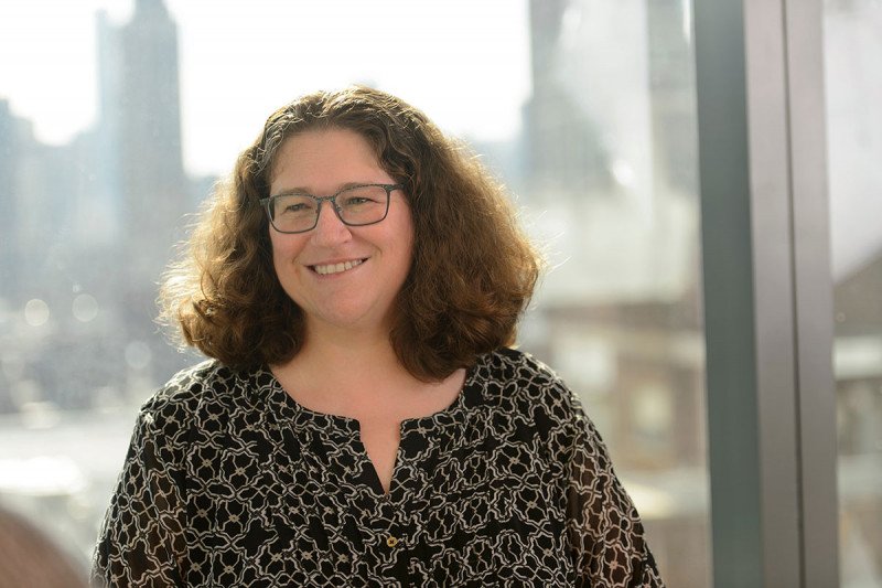
“These studies are focused on efforts to look at tumors at the level of each individual cell,” says Dana Pe’er, Chair of SKI’s Computational and Systems Biology Program and senior author of both papers. “Without going down to the level of single cells, we aren’t fully able to understand what’s going on and what is driving a particular cancer.”
A Growing Field Seeks to Map Cancer
The field of single-cell analysis has expanded greatly in recent years. This is due in large part to rapid technological advances. Mathematical and computational techniques now make it possible to sort the huge quantities of data that are generated by these analyses.
The Human Cell Atlas brings together investigators from all over the world to create a comprehensive reference map of all human cells. Dr. Pe’er, who co-chairs one of this project’s analysis working groups, says the effort has the potential to impact the understanding of many diseases, not just cancer. Collaborative endeavors like this, she notes, can answer fundamental questions about human development.
One of the primary tools in this field is a genomic analysis technique called single-cell ribonucleic acid sequencing (RNA-seq). This system looks at RNA rather than DNA. It enables investigators to determine which genes are expressed, or “turned on,” in particular cells, rather than just which genes are present in the DNA. Because every cell in the body contains the same DNA, RNA analysis provides much-needed detail about cell function and activity.
Characterizing the Immune Landscape of Breast Cancer
One of the new papers is a collaboration among SKI computational biologists and immunologists as well as Memorial Sloan Kettering’s breast cancer team. The study looked at cells taken from human breast tumors. The team also considered normal breast tissue, blood, and lymph node tissue. The investigators analyzed 45,000 immune cells taken from eight tumors and 27,000 additional immune cells.
Identifying such a large number of immune cells could explain why immunotherapy doesn’t always work the same way in each person. Immunotherapy harnesses the power of the body’s natural immune response to fight cancer. Some types of immune cells attack cancer, while others protect tumor cells from harm.
“One of our major findings was that there was a much greater diversity in the states of immune cells found in tumors compared with what we found in normal tissues,” says Alexander Rudensky, Chair of SKI’s Immunology Program and co-senior author of the breast cancer study. A cell’s state is based on not only what type of cell it is but also other factors, such as its location, size, and structure.
“We were surprised to find that, rather than distinct differences between tumor and nontumor tissue, there was a gradient in the levels of different immune cell states,” he says.
In other words, they found a range of differences in the immune makeup of these tissues, not a clear line between the immune cells present in cancer and normal tissue. “This helps explain why tumors have a range of behaviors — not all tumors respond in the same way to immunotherapy,” adds Dr. Rudensky, who is also a Howard Hughes Medical Institute investigator. “But at the same time, we saw common features among the breast cancer samples that were not seen in the normal tissues. From this work, we can start to think about how to develop immunotherapy that’s custom-built for people based on their individual tumor microenvironment.”
Dr. Rudensky explains that the focus on breast cancer was only a starting point to see if the approach would work. The researchers have plans to expand this research to many other types of cancer. “Until very recently, analyzing the data from thousands of cells at the same time would have been a major undertaking,” he says. “But thanks to the transformative work that’s been done by Dr. Pe’er’s team, we can start to grow this area of immunology research.”
Uncovering Hidden Data with MAGIC
The other Cell paper concentrates on identifying the differences among cancer cells themselves, rather than looking at immune cells in their midst. A culmination of five years of work from Dr. Pe’er, the study focuses on cutting-edge ways to reduce the high levels of noise and highlight the biological trends that come from such large quantities of data.
Dr. Pe’er compares the challenge to a common event on crime shows like CSI, in which an image from a photograph is too blurry to make out. “The detectives bring in an expert who has created a computer algorithm to analyze the pixels, allowing them to clearly read the letters and numbers on a license plate,” she says. “In the same way, we have developed a way to reduce the fuzziness in our data and see clear patterns.”
Dr. Pe’er’s collaborators included Smita Krishnaswamy, a former postdoctoral fellow in her lab who now leads her own lab at Yale University. The team dubbed the technique MAGIC for Markov affinity-based graph imputation of cells. The algorithm can recover gene expression data that may be missing from an individual cell if not all of the RNA has been captured.
“Cancer cells have a range of activities. The ones that have the ability to outsmart drugs or to spread to another part of the body are actually quite rare,” she concludes. “Only by looking at the tumor with single-cell resolution are we able to identify them and study them, enabling us to get to the bottom of what gives them these capabilities.”


