Research advances in lymphoma, leukemia, and multiple myeloma are being presented by Memorial Sloan Kettering Cancer Center (MSK) researchers at the American Society of Hematology (ASH) Annual Meeting, which runs December 7–10. Below are some highlights.
Dual-Targeting of JAK and PI3K May Be a Promising Therapeutic Approach for Some T Cell Lymphomas

Dual-targeting of JAK and PI3K may be a promising therapeutic approach for patients with relapsed or refractory peripheral and cutaneous T cell lymphomas, a phase 1 study led by MSK researchers has found.
The trial was broken into two parts to assess the efficacy and safety of dual-targeting of JAK and PI3K with ruxolitinib and duvelisib. Part 1 of the study sought to determine the maximum tolerated dose. Part 2 assessed efficacy based on the presence or absence of JAK/STAT pathway activation. JAK and PI3K are important signaling pathways in cells and play key roles in regulating cell growth, survival, proliferation, and differentiation.
A total of 49 patients were enrolled in the study, with a median age of 65. Most patients had received prior treatments. In the first part of the study, the maximum dose was determined to be 20 mg of ruxolitinib plus 25 mg of duvelisib, twice per day. In the second part, 45% of patients showed a significant improvement on the treatment, with 22% experiencing complete remission, according to findings presented by hematologist-oncologist Alison Moskowitz, MD.
“This is an area of unmet need, and we find these results very encouraging,” Dr. Moskowitz says.
The combination was well-tolerated with manageable toxicities, the study team reported. The toxicity profile was better than with most PI3K inhibitors, noted the team, possibly due to the addition of ruxolitinib. The combination showed particular efficacy in T-follicular helper lymphomas and T-prolymphocytic leukemia, as well as cancers with JAK/STAT activation.
Cross Talk Between Cancer Cells and Immune T Cells May Be Key to Acute Myeloid Leukemia

The immune system’s failure to stop acute myeloid leukemia (AML) from developing may be caused by suppressive signals from abnormal, malignant white blood cells, called leukemia blasts, MSK researchers have found. The signals neutralize the cancer-fighting power of immune T cells.
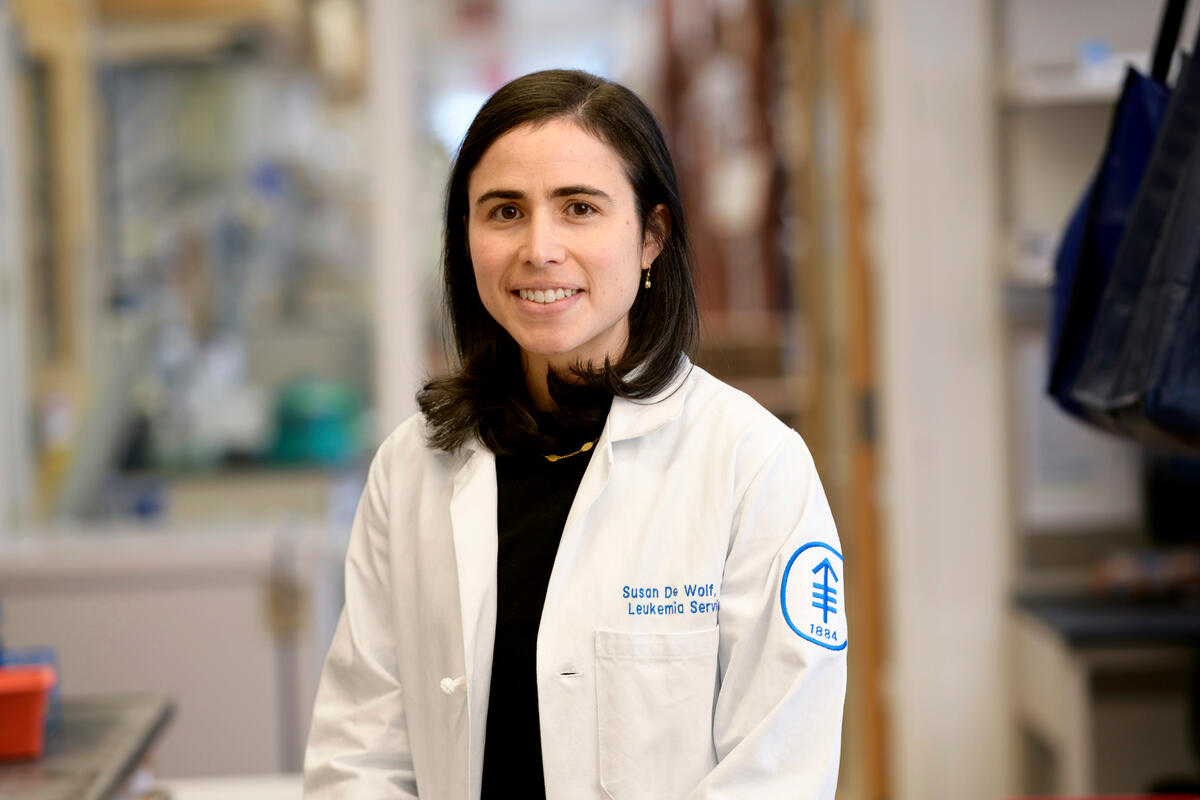
The research team, led by hematologic oncologist Susan De Wolf, MD, also identified multiple surface proteins on both the blasts and the T cells that could be targeted by drugs to boost the immune defense against AML.
“With solid tumors, we talk about the microenvironment of the surrounding cells and tissue and how it affects the immune cells,” Dr. De Wolf says. “With blood cancers like AML, the bone marrow itself is the microenvironment. We wondered: What do these T cells look like given that they’re surrounded by this sea of leukemic blasts, and what could be affecting their behavior?”
The researchers noticed that people with more aggressive AML have large numbers of regulatory T cells (Tregs) in the bone marrow. These cells tamp down the immune response to prevent the body from attacking itself. But there also are CD8 T cells present. These immune cells have cancer-killing ability but are strangely ineffective.
Working with computational oncologist Sohrab Shah, PhD, the researchers were able to track the gene expression of the CD8 T cells in the presence of AML. The CD8 T cells appeared to lose their cancer-killing ability over time, suggesting that the microenvironment — specifically signals from the leukemia blasts — were disarming them.
In a mouse model for AML, the researchers then showed that removing the Tregs enabled the CD8 T cells to regain their cancer-killing activity and wipe out the disease. The scientists think that cross talk between the blasts, the CD8 cells, and the Tregs helps the cancer cells avoid the immune attack. The CD8 cells are stopped from attacking, and Treg cells are recruited to the bone marrow to further suppress the immune response.
“This is the largest, most in-depth study characterizing what is happening in the leukemia microenvironment,” Dr. De Wolf says. “This really opens up new avenues for testing therapies.”
Whole-Genome Sequencing Reveals Some Differences in Multiple Myeloma Among Patients With African Ancestry

Some types of cancer occur more frequently in people with particular backgrounds, due to either genes linked to a person’s ancestry or to environmental factors. One cancer where incidence varies across population groups is multiple myeloma, a type of blood cancer that affects cells in the bone marrow called plasma cells. People of African ancestry appear more likely to develop multiple myeloma than other groups.
At the ASH meeting, a multicenter team of investigators led by MSK myeloma specialist Kylee Maclachlan, MBBCh, PhD, reported results from the analysis of DNA from 340 patients with either multiple myeloma or a precancerous condition that often leads to myeloma. About 26% of the patients had African ancestry. The DNA was studied using whole-genome sequencing, a comprehensive way to look at all the genes found in a person or organism.
The investigators discovered that most genetic features of the cancer were similar across ancestry groups. However, people with African ancestry, despite having a higher genetic risk score for the disease, had lower overall number of mutations. They were also more likely to have a simpler type of myeloma and were found to have less activity from a family of proteins called APOBEC, which are linked to some DNA changes related to cancer. By contrast, people of European ancestry had more APOBEC activity and more complex disease. “This research suggests that, with comparable treatment, patients with African ancestry would be expected to have better clinical outcomes than a comparable group with European ancestry,” Dr. Maclachlan says.
This work was done in collaboration with the nonprofit New York Genome Center, of which MSK is a founding member, and its Polyethnic-1000 consortium. This project aims to harness New York’s diverse populations to understand the influence of different genetic backgrounds on cancer formation and growth.


