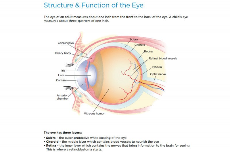
The cornea is the clear portion of the front of the eye. The conjunctiva is a tissue which lines the eyelids and the eyeball up to the edge of the cornea. The iris is the colored portion of the eye. It is made up of a spongy tissue and is an extension of the choroid. The pupil is the opening in the iris which allows light into the eye. The lens helps focus light rays onto the retina. The lens can change shape, or “accommodate,” to focus on near or distant objects.
The eye is filled with fluids which help nourish and maintain the pressure within the eye. The anterior chamber, the front portion of the eye between the iris and the cornea, is filled with aqueous humor, a watery fluid, which nourishes the lens and maintains the pressure within the eye. The back portion of the eye is filled with vitreous humor, a transparent gel. The retina is made up of ten layers and contains millions of cells. The optic nerve has nerve fibers which transmit information to the brain for interpretation of objects seen and contains about a million cells.
Areas of the Eye Responsible for Vision
The macula is the area of the retina that is responsible for central vision. Its central portion is referred to as the fovea and is responsible for the sharpest vision. The macula houses the highest concentration of the cones which are responsible for color and sharp vision. The rest of the retina is composed of rods, which are more sensitive to light and are responsible for night vision and peripheral vision.
Eye Muscles
Attached to the outside of the wall of the eye are six muscles that aid in the movement of the eye. Movement of the eye is caused by shortening of the eye muscles.

