By Ian Demsky
The field of oncology hums with data. Imaging scans, laboratory tests, tumor mutations, medications and dosage — they’re all captured in electronic record systems.
There’s a clinical record of how a person was treated and how the cancer they had responded to the treatment, of what worked and for how long.
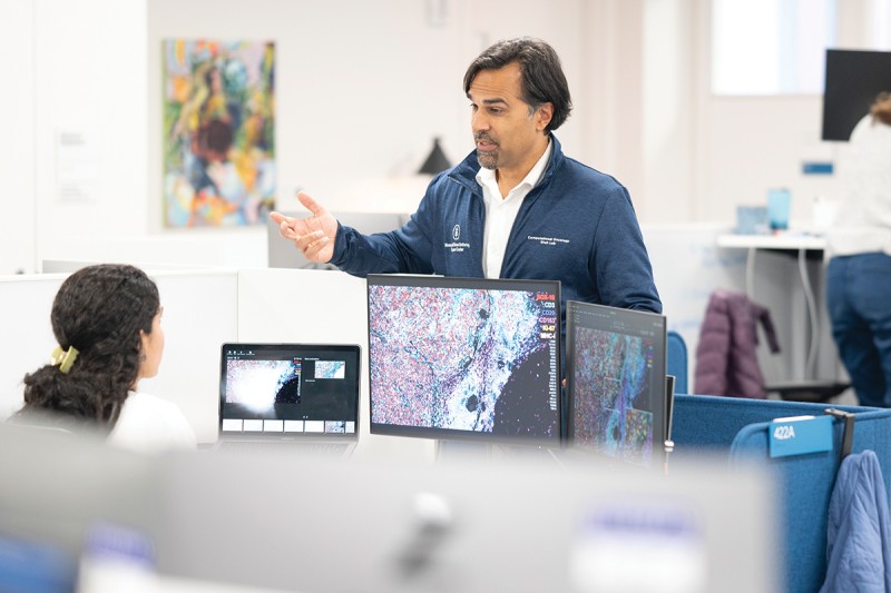
“The primary purpose of gathering all this information is to treat each patient properly and effectively,” says Sohrab Shah, PhD, who heads the Computational Oncology Program at Memorial Sloan Kettering Cancer Center (MSK). “But all this information has enduring value by creating a large data set from which patterns can be analyzed for the benefit of future patients.”
Away from the clinic, too, across MSK’s 100-plus research laboratories, sophisticated computational methods are playing an increasingly vital part in answering fundamental questions about human biology and cancer biology. Here, scientists from “wet labs” (think white coats, petri dishes, and microscopes) team up with specialists from “dry labs” (think computer models and statistics) to decipher the troves of data generated by modern research technologies.

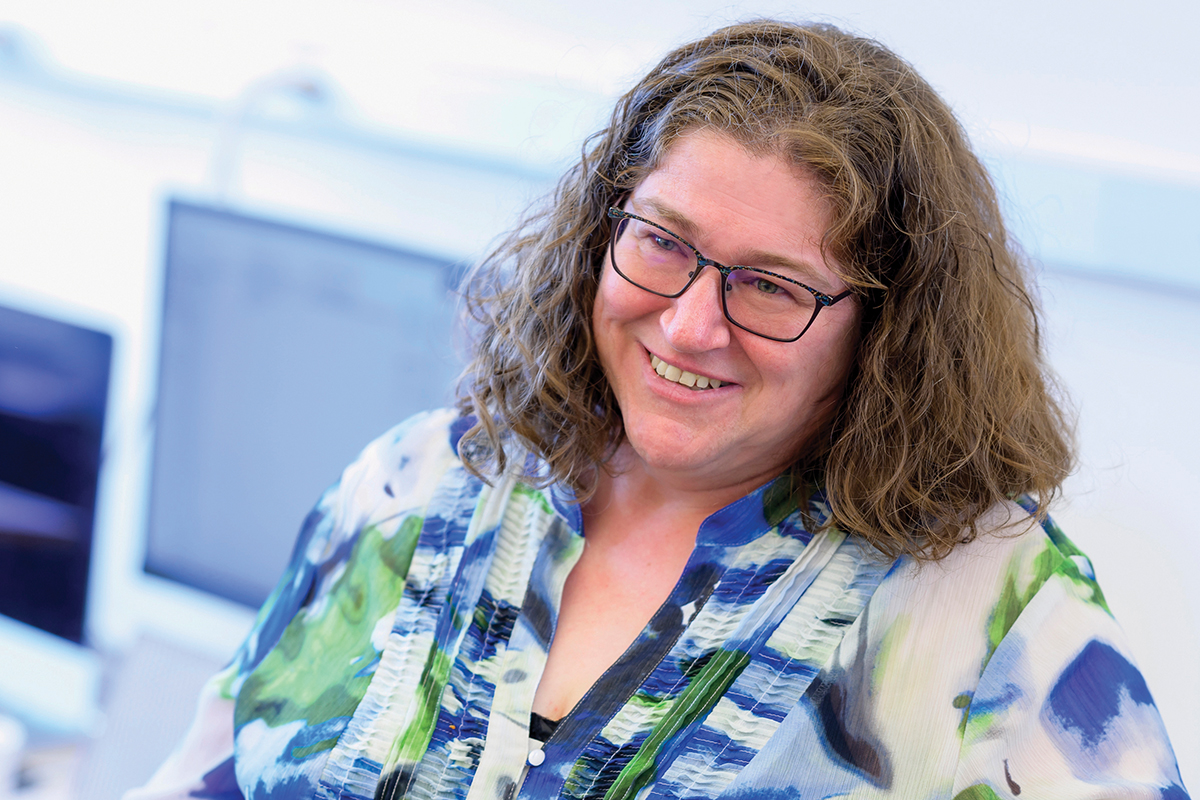
“Biology is really becoming an information science,” says Dana Pe’er, PhD, Chair of the Computational and Systems Biology Program at MSK’s Sloan Kettering Institute. And what sets MSK apart is the ability for computational and cancer experts to work together as partners.
“You can’t just blindly swing the latest computational method at a problem, out of the box. It doesn’t work,” says Dr. Pe’er, who is also a Howard Hughes Medical Institute Investigator, one of the highest recognitions in science. “You have to model the problem based on the right assumptions. And for that, biological expertise is indispensable.”
Teaching Machines To Recognize Cancer
One powerful tool for scientists and doctors is artificial intelligence (AI). The technology has been in the spotlight in recent months as AI chatbots and image generators have become popular, leading to renewed conversations about its role in medicine.
AI’s most immediate application for patient care is to help humans pore over digital pictures — such as diagnostic images and pathology slides. These are promising tools with the potential to significantly augment a human expert’s perception, stamina, and efficiency. But, on the whole, they are still being fine-tuned.
“Machine learning is very good at what you’ve taught it,” Larry Norton, MD, Medical Director of MSK’s Evelyn H. Lauder Breast Center, told Good Morning America during a segment on the increasing use of AI to help radiologists detect breast cancer. “But when machines see something they have no experience with, they’re not very good at identifying it.”
And while AI technology is getting better all the time, it is not yet considered the standard of care, Dr. Norton notes. “A skillful radiologist is still your best partner,” he says. “And your best protection is getting screened — about half of people who should be getting annual mammograms are not getting them.”
What’s critical, Dr. Pe’er says, is for experts to work together on each type of clinical task. “There are cases when the AI can perform much better than a pathologist alone, but this can only happen with the right training,” she says.
Enlisting AI in Cancer Research
Meanwhile, AI has had a quiet and notable role in bioscience research for years, decades even, Dr. Shah notes.
For example, Dr. Shah and MSK pathologist Jennifer Sauter, MD, were co-senior authors of a recent study that used AI to combine data from patients’ digital pathology slides and CT scans to better predict immunotherapy outcomes. Real-world data was used to teach the program to find patterns that could predict whether a given person’s lung cancer would be likely to respond well to immunotherapy.
Immunotherapy, of course, has been revolutionary for many patients, especially those with lung cancer — but it doesn’t work for a large number of people. So researchers continue to look for ways to predict who will be likely to benefit from immunotherapy, as well as for ways to make treatment work better for more people.
“Our method outperformed standard-of-care approaches considerably by combining different sources of data,” Dr. Shah says. “And this type of expert-guided machine-learning approach holds a lot of promise for improving our ability to provide personalized care.”
Meanwhile, Dr. Pe’er points to a collaboration with neuro-oncologist Adrienne Boire, MD, PhD, where computational methods helped figure out how cancer cells survive in the barren environment of the cerebrospinal fluid.
Using single-cell RNA sequencing — which requires high-octane math and statistics to sort through huge quantities of data about gene activity across tumor cells — the researchers were able to show that iron-hungry cancer cells reprogram themselves to gobble up all the nearby iron. The discovery suggested a new treatment approach, and the findings formed the basis for a phase 1 clinical trial that launched in 2022.
“Oftentimes, there are so many factors and data points that come into play in biology, it’s well beyond our human ability to perceive meaningful patterns,” Dr. Pe’er says. “Still, it takes both computational and biological expertise to frame the questions in the right way for a computer to find sensible and actionable patterns. And as computational cancer researchers, that’s our craft.”
Dr. Boire holds the Geoffrey Beene Junior Faculty Chair. Dr. Norton holds the Norna S. Sarofim Chair in Clinical Oncology. Dr. Pe’er holds the Alan and Sandra Gerry Endowed Chair. Dr. Shah holds the Nicholls-Biondi Chair.